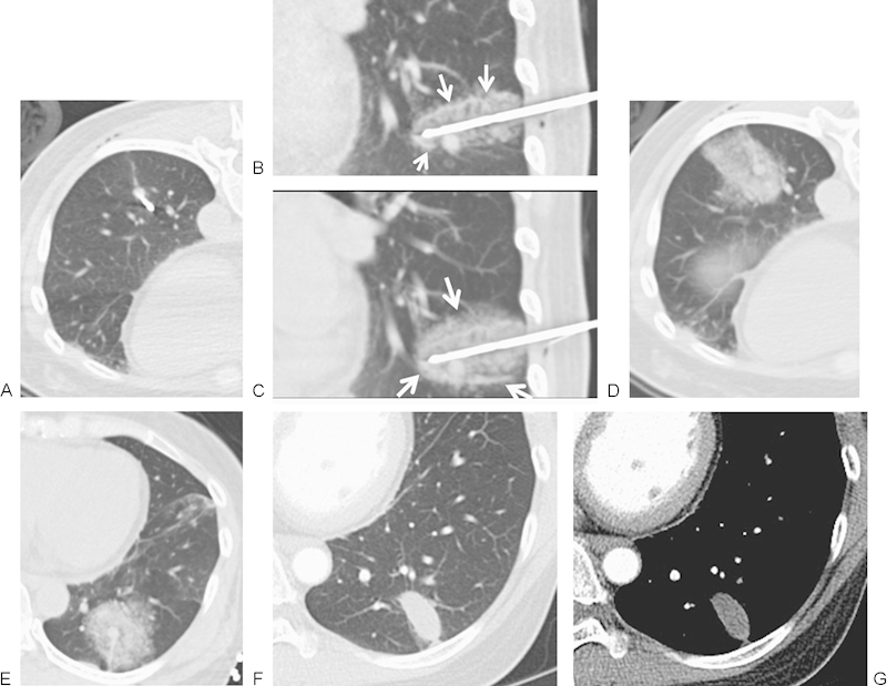
Figure 5.

(A) A 50-year-old woman with solitary metastatic colon carcinoma to left lower lobe. Multiple attempts were made to place the probe through the nodule; however, the nodule changed position, a common finding while placing larger cryoprobes in smaller subcentimeter nodules. Ultimately the cryoprobe was placed at the superior surface of the nodule. (B) Initial 3 minutes of freeze cycle followed by passive thaw was performed to create hemorrhage surrounding the nodule and to allow for the definite freeze cycle. Peripheral rim of hyperdensity represents the edge of the melting ice ball (arrows), and the inner hypodensity surrounding the probe represents the ice ball itself. (C) Following a second freeze cycle of 10 minutes, the edge of the ice ball is seen (arrow) extending beyond the initial hemorrhage. (D) Immediately postablation, there is development of hemorrhage at the ablation site representing necrosis and hemorrhage. (E) The patient was placed in a supine position to limit the transbronchial spread of hemorrhage. (F, G) At 1 week following cryoablation, computed tomography demonstrates an elliptical ablation zone with resolution of hemorrhage. The size of the ablation zone is larger than the ablated nodule and depends on the number and size of the original ablation probes.
