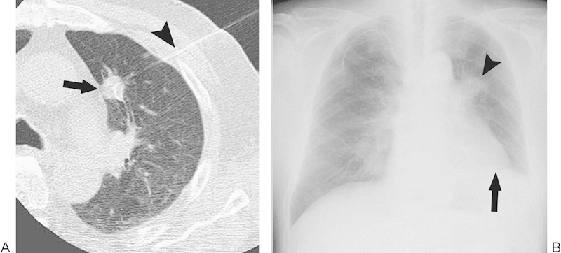
Figure 4.

Phrenic nerve injury. (A) Computed tomography image during radiofrequency ablation (RFA) show a multi-tined expandable electrode (arrowhead) introduced into a tumor (arrow) close to the pulmonary trunk. (B) Chest radiograph after RFA demonstrates elevation of the diaphragm on the treated side (arrow). The arrowhead denotes the ablation zone.
