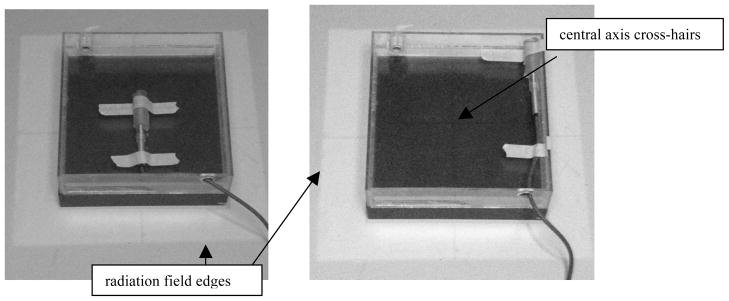
FIG. 2.

A calibrated ion chamber was used to verify the dosimetry on (left) and off (right) the central axis. The illuminated light field surrounding the chamber (representative of the radiation field) and the central axis cross-hairs (used for positioning) can be seen.
