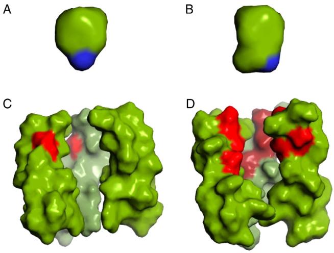
Fig. 2.
Comparison of the amphipathic drugs, amantadine (A) and rimantadine (B), with the pores from AM2 (C, PDB 2H95) and BM2 (D, PDB 2KIX) on the same scale. The AM2 and BM2 structures are represented by only 3 of the 4 helices so that a view into the pore can be achieved. Furthermore, only residues Leu26-Ile42 (AM2 numbering) are displayed. The amino group of the drugs is colored blue, the AM2 drug binding site is primarily hydrophobic indicated by green (left) while BM2 (right) channel pore is lined with polar residues in red.

