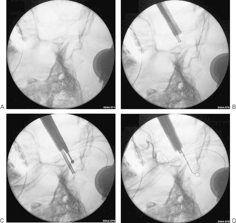
Figure 2.

(A) Preoperative lateral fluoroscopy (C-arm) for depiction of the sella region. (B, C) Intraoperative lateral fluoroscopy for rapid orientation during opening the sphenoid floor. (D) Lateral fluoroscopy for orientation at the final removal of the lesion.
