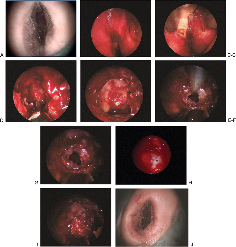
Figure 3.

(A) Nasal inspection at the beginning of the procedure. (B) Localization of the sphenoid ostium at the recessus spheno-ethmoideus. (C) View at the floor of the sphenoid cavity after breaking the septum. (D) Removal of the sphenoid floor with preservation of a large bone flap. (E) View at the sphenoid cavity with a septum at the right (+), the carotid channel (*) at the left and the floor of the sella. (F) Tumor removal under endoscopic view. (G) Final endoscopic inspection. In the depth the diaphragm. (H) Inspection of the sellar region with 30 degree optic. At the right the carotid channel (*). No tumor remnants visible. (I) Closure of the sellar with fibrin glue and reconstruction of the sphenoid floor. (J) Postoperatively undamaged nose.
