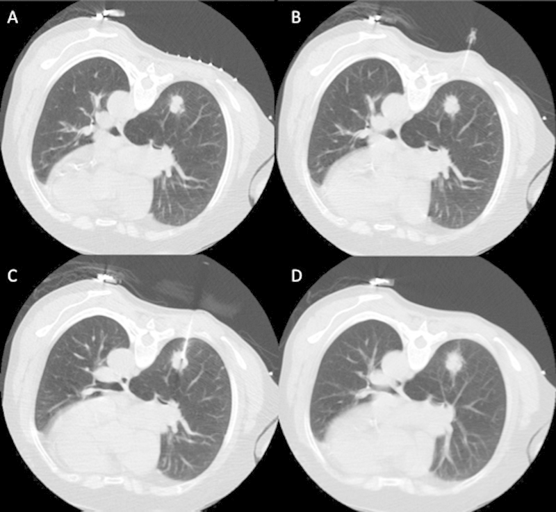
Figure 2.

(A) Example of a normal lung biopsy in a patient with a right lower lobe lung nodule showing the preprocedure scan with a grid on the skin. (B) A 25-gauge needle in the subcutaneous tissues following lidocaine injection. (C) The outer cannula of a coaxial biopsy device within the lesion. (D) A postbiopsy image through the region of biopsy.
