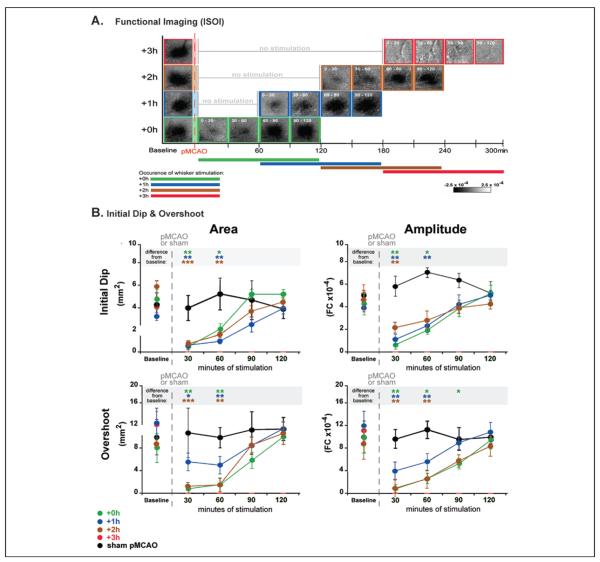
Figure 5.
Evoked functional response returns gradually on stimulation onset in all protected animals throughout the treatment period. (A) Following permanent middle cerebral artery occlusion (pMCAO), a return of cortical function is evident in +0h, +1h, and +2h groups during protective stimulation treatment, and a loss of all three phases of the evoked function response in animals that receive treatment 3 hours post-occlusion (+3h animals). Representative data from functional imaging of the initial dip for each group (+0h, +1h, +2h, and +3h) were arranged according to minutes of treatment delivered (treatment time is included as white font in the top left corner of each image). Groups +0h, +1h, and +2h all regained evoked functional response comparable to baseline after 90 minutes of whisker stimulation, whereas +3h animals never demonstrated any post-pMCAO cortical activity. (B) In each plot, group baseline is plotted with 120 minutes of post-occlusion stimulation period data. Means and standard errors are provided for the area (left) and amplitude (right) of the ipsi-ischemic C2 initial dip (top) and overshoot (bottom) phases of evoked functional response to whisker stimulation before and after pMCAO. +3h animals had no response to stimulation at any time point post-pMCAO. For all other groups (sham-pMCAO, +0h, +1h, and +2h), asterisks within the gray band above each plot indicate a significant difference from baseline, *P < .05, **P < .01, and ***P < .001. Data in panels A and B are from Lay and others (2011b); used with permission of authors and publisher.

