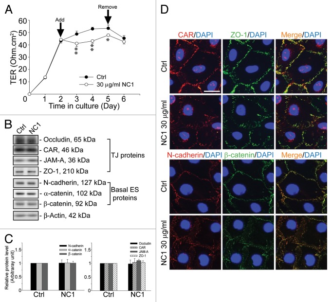
Figure 4. Disruption of the Sertoli cell TJ-permeability barrier function by recombinant Colα3(IV)NC1 domain protein mediated via changes in protein distribution and/or localization at the Sertoli cell-cell interface. (A) Sertoli cells were plated at 1.2 × 10⁶ cells/cm2 on Matrigel-coated bicameral units with an established TJ-permeability barrier by day 2, and 30 µg/ml recombinant Colα3(IV) NC1 (1 µM) in PBS was included in the F12/DMEM. The presence of the NC1 domain recombinant protein was found to significantly perturb the TJ-barrier function vs. PBS control (Ctrl) until the recombinant protein was removed on day 5, and the partially disrupted TJ-barrier was “resealed.” *, p < 0.05; **, p < 0.01. (B) Sertoli cells were plated at 0.5 × 10⁶ cells/cm2 for 2 d before 30 µg/ml (1 μM) of the recombinant NC1 domain was included in the F12/DMEM. Two d later, there was no significant change in steady-state levels of TJ and basal ES proteins when compared with PBS control (Ctrl). (C) This histogram summarizes results shown in (B) where protein levels from Ctrl were arbitrarily set at 1. Each bar is a mean ± SD of n = three independent experiments. (D) Sertoli cells were cultured at 0.045 × 10⁶ cells/cm2 on Matrigel-coated coverslips, and treated with 30 µg/ml NC1 or PBS Ctrl for 2 d. Cells were fixed and stained for CAR (red) and ZO-1 (green) or N-cadherin (red) and β-catenin (green) by dual-labeled immunofluorescence analysis using the corresponding primary antibodies and Alexa Fluor 555- or 488-conjugated secondary antibodies (see Table 1). Sertoli cell nuclei were visualized by DAPI (blue). CAR, ZO-1, N-cadherin and β-catenin were all seen at the Sertoli cell-cell interface in Ctrl cells while treatment of cells with 30 µg/ml NC1 led to changes in distribution of these BTB-associated cell adhesion proteins, moving from the cell-cell interface into the cell cytosol. Scale bar, 5 µm, which applies to all micrographs. Findings of this dual-labeled immunofluorescence analysis experiment reported herein were results of a representative experiment, which was repeated three times using different Sertoli cell cultures and yielded similar results.

An official website of the United States government
Here's how you know
Official websites use .gov
A
.gov website belongs to an official
government organization in the United States.
Secure .gov websites use HTTPS
A lock (
) or https:// means you've safely
connected to the .gov website. Share sensitive
information only on official, secure websites.
