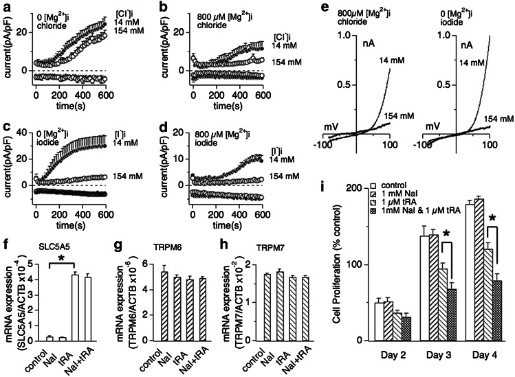
Fig. 5.
Extracellular iodide suppresses breast cancer cell proliferation in dependence of SLC5A5 sodium-iodide symporter expression. a Whole cell currents were recorded in MCF-7 breast cancer cells and normalized to cell capacitance (in pF). Cells were perfused with 14 or 154 mM Cl− in the absence of intracellular Mg2+ (n = 6–8). b Cells were perfused with 14 or 154 mM Cl− in the presence of 800 μM free Mg2+ in the internal solution (n = 6–8). c Low (14 mM) or high (154 mM) iodide perfusion of MCF-7 in the absence of intracellular Mg2+ (n = 6–8). d Low (14 mM) or high (154 mM) iodide perfusion of MCF-7 with additional 800 μM free Mg2+ (n = 6–8). e Representative current voltage (I/V) curves for native TRPM7 currents in MCF-7 cells in the presence of intracellular 14 and 154 mM halide ions (Cl− or I−) described in panel b or panel c, respectively. All I/V curves were extracted at 600 s whole-cell time and were derived from a high-resolution current record in response to a voltage ramp of 50 ms ranging from −100 to +100 mV. f, g, h Changes of mRNA expression of SLC5A5 (sodium-iodide symporter, panel f), TRPM6 (panel g) and TRPM7 (panel h) by treatment of 1 mM sodium iodide (NaI), 1 μM trans-retinoic acid (tRA) or a combination thereof. The expression of each mRNA is normalized to β-actin expression. Data are mean ± SEM from three independent experiments. Note that exposure of MCF-7 cells to 1 μM tRA, but not 1 mM NaI, significantly increases SLC5A5 mRNA expression. The same treatment had no significant effect on TRPM6 (panel g) or TRPM7 mRNA expression (panel h). i Effect of NaI on MCF-7 breast cancer cell proliferation. The histogram shows the mean number of viable MCF-7 cells counted on the 2nd, 3rd and 4th day after cell seeding. Cells were planted at a density of 1.5 × 10⁵ ml−1 with the medium containing 10 % FBS. The cells were then treated with medium vehicle (control), 1 μM tRA, 1 mM NaI or 1 μM tRA and 1 mM NaI combined (n = 4 independent experiments). Error bars indicate SEM. Star indicates P < 0.002

