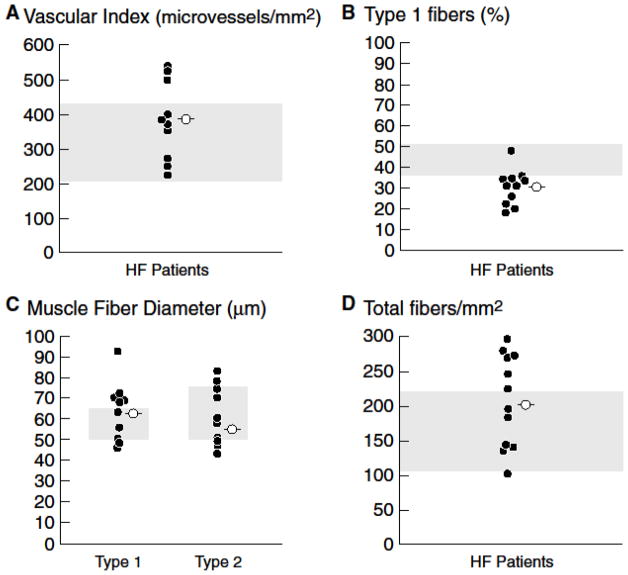
Figure 2.
Scatter plots of individual measures (filled circles) and mean values (open circles) of morphometry in 11 HF patients. Panel A. Vascular index was within or above the normal range in all HF patients. Panel B. The proportion of type 1 fibers was diminished in 10 of 11 HF patients, consistent with a fiber shift from oxidative type 1 fibers to glycolytic type 2 fibers. Panel C. The overall mean fiber diameter is within the normal range in HF patients on optimal medical and device therapy. Panel D. Total number of fibers per mm2 is within the normal range in HF patients on optimal medical and device therapy. Shaded area represents the normal range established in prior published studies from the UCLA Neuropathology Lab [26] and in the literature [25,31,32].

