


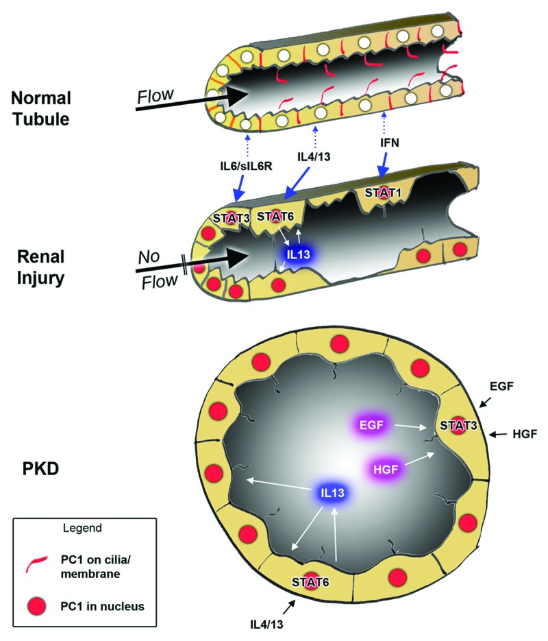
Figure 2. Model on how the cleaved, nuclear PC1 tail can lead to sensitization to STAT-activating growth factors. In normal renal tubule cells, PC1 expression is low and its localization to restricted to primary cilia and cell junctions (red). Renal injury leads to increased PC1 expression, cleavage of its cytoplasmic tail that then traffics to the nuclei (red) where it can co-activate STAT transcription factors. Depending on the growth factor/cytokine environment, different STATs will be activated. In injured tubules, this will lead to amplified STAT activity whereas intact tubules should be relatively insensitive (due to lack of nuclear PC1 tail). Different cytokines will lead to different responses. For example, IL13 secretion by M2 macrophages would cause STAT6 activation in tubule cells which may activate tissue regeneration. In contrast, interferon secretion by M1 macrophages may trigger cell death by activation of STAT1 in damaged tubule cells. In PKD, overexpression of (mutated) PC1 may lead to constitutively high levels of the nuclear PC1 tail which will hyper-sensitize tubule cells to cytokine signaling leading to inappropriate responses. Because factors secreted into cyst lumens cannot escape, positive feedback loops establish themselves which lead to persistent activation of STAT6 and STAT3.