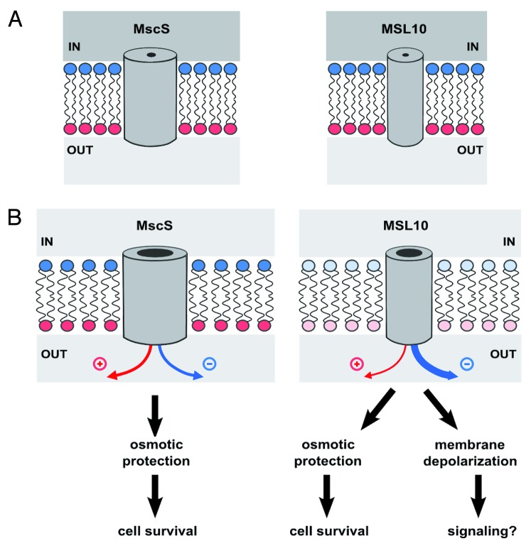
Figure 3. Model for the function of MSL10, as compared with MscS. (A) MscS and MSL10 channels in cells under severe hypoosmotic stress, before the channels open. Channels are drawn as simple tubes piercing the lipid bilayer. Positively and negatively charged monolayers are indicated with red and blue lipid headgroups, respectively. Hyperosmotic medium inside the cell is dark gray, while hypoosmotic medium in the extracellular space in light gray. (B) After opening of the channels. Both MscS and MSL10 release osmolytes, providing protection from lysis and allowing cell survival. However, the increased preference for anions of MSL10 results in the depolarization of the plasma membrane upon gating (shown as decreased intensity of red and blue). Therefore, MSL10 functions are likely not restricted to relief of hypoosmotic stress but also may include signaling through downstream depolarization-activated ion channels.

An official website of the United States government
Here's how you know
Official websites use .gov
A
.gov website belongs to an official
government organization in the United States.
Secure .gov websites use HTTPS
A lock (
) or https:// means you've safely
connected to the .gov website. Share sensitive
information only on official, secure websites.
