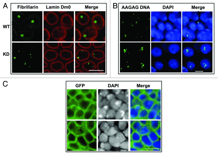
Figure 4. AAGAG/CUCUU RNA knockdown lead to nuclear defects. (A) AAGAG/CUCUU KD embryos show multiple nucleoli. WT and KD embryos were immunostained for fibrillarin and lamin Dm0. WT nuclei show single nucleolus, whereas nuclei in KD embryos show multiple nucleoli. No change was detected in lamin Dm0 staining. Scale bar, 10 μm. (B) Localization of satellite DNA is disturbed in nuclei of wing imaginal discs of AAGAG-knockdown larvae. DNA-FISH with WT and KD nuclei was done using DIG-labeled AAGAG probe. DNA was visualized by DAPI. In WT nuclei, satellite DNA was seen as one or two condensed spots close to the nuclear periphery, while in KD it moves toward interior of the nuclei and was split into many foci. Scale bar, 5 μm. (C) AAGAG-knockdown nuclei show defective chromatin packaging. In this experiment, RNAi was induced in localized areas of wing imaginal disc using omb-Gal4 driver. GFP positive cells mark the area where RNAi is executed. KD wing imaginal discs show disturbed nuclear DAPI staining where heterochromatin was spread out and diffused. WT discs showed condensed heterochromatic spots in equivalent positions. Scale bar, 5 μm.

An official website of the United States government
Here's how you know
Official websites use .gov
A
.gov website belongs to an official
government organization in the United States.
Secure .gov websites use HTTPS
A lock (
) or https:// means you've safely
connected to the .gov website. Share sensitive
information only on official, secure websites.
