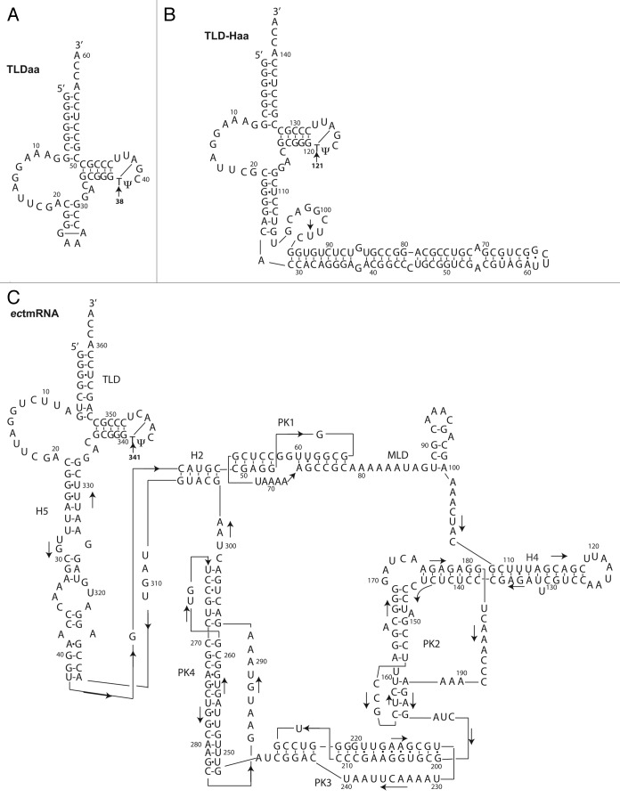
Figure 1. Secondary structure models of (A) TLDaa, (B) TLD-Haa and (C) full-length ectmRNA. The TLD parts of the A. aeolicus and E. coli models are very similar in sequence. An arrow stresses the position of the ribothymidine m5U (labeled T for simplification) (i.e. 38 in TLDaa, 121 in TLD-Haa and 341 in ectmRNA). The second methylated uridine observed in TLD-Haa, as explained in text, is most probably U97.

An official website of the United States government
Here's how you know
Official websites use .gov
A
.gov website belongs to an official
government organization in the United States.
Secure .gov websites use HTTPS
A lock (
) or https:// means you've safely
connected to the .gov website. Share sensitive
information only on official, secure websites.
