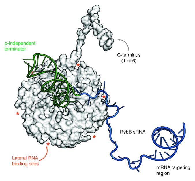
Figure 3. Model of an Hfq/sRNA complex. Proximal side view of a model of the Hfq/RybB complex. The Hfq hexamer (PDB-ID: 2YLC9) is shown as a surface representation with a superimposed model of only one of the six Hfq C-termini for clarity. RybB sRNA is depicted in cartoon representation with the ρ-independent terminator colored in green, the single-stranded sequence is blue and the location of the seed region is indicated. The asterisks mark the location of the six lateral RNA binding sites of Hfq. The model was assembled using COOT98 considering the biochemical and structural evidence summarized in this review. The depicted structure of the C terminus was modeled using HHpred.99 The model shows how Hfq might interact with RybB sRNA and also gives an impression of the proportions of the sRNA body with respect to the size of the terminator stem-loop and the Hfq protein.
