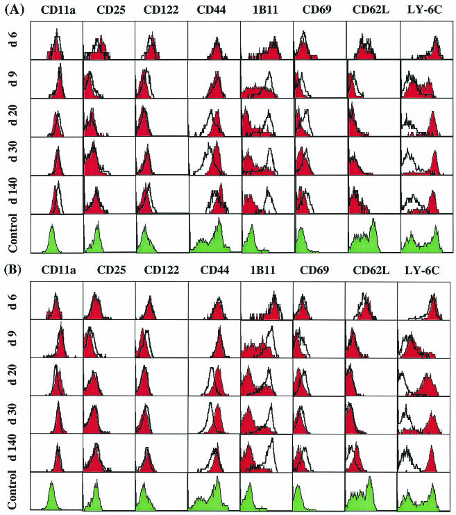
FIG. 9.
Phenotypic analysis of virus-specific CD8+ T cells from the spleen and liver during acute versus persistent infections. Lymphocytes isolated from the spleens (A) or livers (B) of C57BL/6 mice infected with 102 PFU (red histograms) or 2 × 10⁶ PFU (open, thickly lined histograms) of LCMV-Docile at the indicated times after infection were triple stained with anti-CD8α, an antibody specific for CD11a (LFA-1), CD25 (IL-2-Rα), CD122 (IL-2-Rβ), CD44, 1B11 (CD43), CD69, CD62L, or Ly-6C, and the Db/GP133-41 tetramer. As a control, cells from uninfected mice were double stained for CD8α and the activation markers listed above (green histograms). Histograms are gated on cells that were positive for CD8α and Db/GP133-41 tetramer. These results are representative of three separate experiments.

