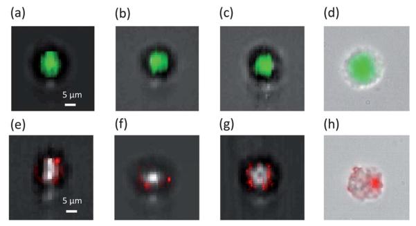
Fig. 5.
Fluorescence images of stained HeLa cells collected by FOFM. (a–c) The fluorescence images of Acridine Orange stained HeLa cells acquired by FZP FOFM superimposed on defocused bright field SROFM images to provide the outline. (e–g) The fluorescence images of Qtracker labled HeLa cells acquired by FZP FOFM superimposed on defocused bright field SROFM images. (d,h) The fluorescence image superimposed on a bright field image acquired by a conventional 20X 0.4 N.A. microscope.

