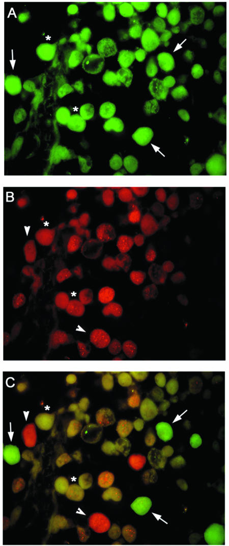
FIG. 4.
Double-labeling immunofluorescence for JCV T antigen and EBV LMP1 in primary CNS lymphoma cells. Samples of primary CNS lymphoma were evaluated by double immunolabeling for the presence of JCV T antigen (A) and EBV LMP1 (B). (C) Superimposition of panels A and B in which JCV T-antigen-positive cells are green and LMP1-positive cells are red. Representative tumor cells expressing T antigen alone are indicated with an arrow; arrowheads indicate cells expressing LMP1 only, and cells positive for both T antigen and LMP1 are indicated with an asterisk. Magnification, ×1,000.

