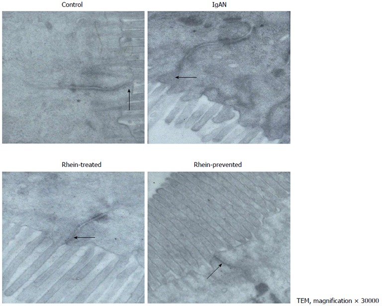
Figure 1.

Electron micrograph of intestinal epithelial cells showing tight junction. A: In the control group, the tight junction appeared as an electron-dense belt at the apex of the intestinal epithelial cells (arrow), indicating an intact intestinal mucosal barrie; B: In the IgA nephropathy (IgAN) group group, the intercellular space was widened, the tight junction was indistinct, and the density was reduced (arrow); C and D: In the Rhein-treated and Rhein-prevented groups, the density of the tight junctions was increased compared with that in the IgAN group (arrows). TEM: Transmission electron microscopy.
