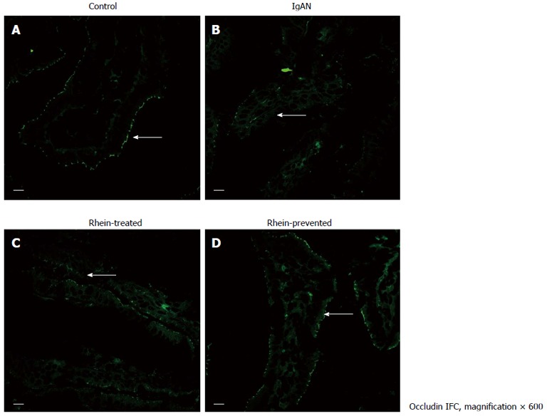
Figure 2.

Location of tight junction protein occludin in rat ileum. Laser confocal microscope immunofluorescence staining of ileum from all four groups of rats. A: Cross-section of a normal intestinal villus. Immunoreactive occludin was localized at the apex of intestinal epithelial cells, consistent with the site of the intestinal mucosal barrier (arrow); B: Cross-section of an intestinal villus in the IgA nephropathy (IgAN) group. Occludin immunofluorescence staining became weak and discontinuous (arrow); C and D: Cross-section of an intestinal villus in the Rhein-treated group and cross-section of an intestinal villus in the Rhein-prevented group. Compared with the IgAN group, occludin immunofluorescence staining became stronger and continuous (arrows). Scale bars = 10 μm. IFC: Integrated fluidic circuit.
