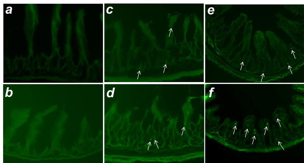
Figure 1. MSC intestinal engraftment.
Shown are representative images of: a) uninjured jejunum from an animal subjected to sMAO and treated with AF-MSC, b) terminal ileum from a sham operated animal treated with AF-MSC, c) terminal ileum from an animal subjected to sMAO + BM-MSC, d) terminal ileum from an animal subjected to sMAO + BM-MSC + HB-EGF, e) terminal ileum from an animal subjected to sMAO + AF-MSC, and f) terminal ileum from an animal subjected to sMAO + AF-MSC + HB-EGF. Intestines were viewed with fluorescent microscopy at 200× magnification. White arrows indicate engrafted MSC.

