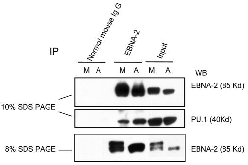
FIG. 5.
Decreased association of PU.1 and EBNA-2 in M-phase-arrested cells when EBNA-2 is hyperphosphorylated. Immunoprecipitation of EBNA-2 was performed with whole-cell lysates from asynchronously growing (A) and nocodazole-arrested M-phase cells (M). A portion of the immunoprecipitated lysates was separated on a 10% gel, and immunoblotting of EBNA-2 and PU.1 was carried out on the same membrane (top and middle panels). The same samples were separated on an 8% gel to show more clearly the EBNA-2 hyperphosphorylation (bottom panel).

