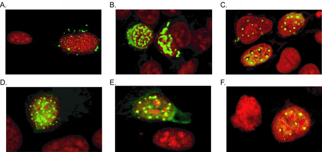
FIG. 2.
Subcellular localization of pIX variants. The subcellular localization of the various pIX proteins was tested by immunofluorescence microscopy. 911 cells grown on coverslips were transfected with the various pIX.RGD expression plasmids. At 2 days postinfection, the localization of pIX was visualized with anti-pIX and FITC-labeled goat anti-rabbit antibodies. The nuclei were stained with propidium iodide. (A) wt.pIX; (B) pIX.RGD; (C) pIX.flag.RGD (D); pIX.flag.30.RGD; (E) pIX.flag.45.RGD; (F) pIX.flag.75.RGD. All pIX variants are localized in nuclear aggregates, as is wt.pIX.

