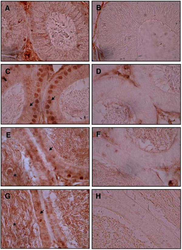
Figure 6.
Immunohistochemistry analyses of SPAG11A in the four regions of the mouse epididymis. The epididymis was isolated, fixed in 4% paraformaldehyde and cut transversely in 5 μm-thick sections. An antibody against human SPAG11A was used to examine the expression of SPAG11A protein in the four epididymal regions. (A) SPAG11A was not present in the initial segment with no staining was detected in the nucleus and cytoplasm of the epithelial cells. (C) SPAG11A was detected both in the nucleus and cytoplasm of the principal cells of the caput. (E and G) Cytoplasmic staining was also detected in the corpus and cauda, respectively. Positive staining was indicated by arrows. (B, D, F, H) Specificity of staining in each region was assessed by comparing with negative controls for initial segment, caput, corpus and cauda, respectively. The negative controls were treated in the same manner as the other samples except PBS was used instead of the primary antibody against SPAG11A. It is interesting to note that positive staining was also detected in the sperm cells inside the lumen of corpus and cauda (*). The images were observed at 1000× magnification.

