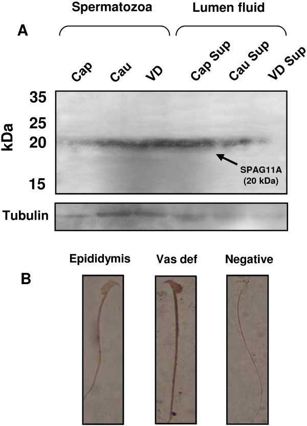
Figure 8.
Western blots and immunocytochemistry revealed the presence of SPAG11A in mouse spermatozoa and epididymal luminal fluid. (A) Protein was extracted from mouse spermatozoa isolated from the caput epididymis (Cap), cauda (Cau) and vas deferens (VD) and luminal fluid from the caput (Caput supernatant, Cap Sup), cauda fluid (Cau Sup) and vas deferens fluid (VD Sup). Fifteen micrograms of protein was separated by 10% SDS-PAGE and transferred to PVDF membranes. The target protein was detected with a rabbit polyclonal anti-human SPAG11A antibody. The results revealed that SPAG11A (20 kDa, indicated by an arrow) was detected weakly in sperm isolated from the caput region, but more SPAG11A was present in cauda and vas deferens sperm. In the luminal fluid, SPAG11A was detected strongly in the caput, reduced towards the cauda and no signal was detected in the vas deferens. The membrane was subsequently stripped and re-probed with an antibody against mouse alpha tubulin as a loading control. (B) Immunocytochemistry on the mouse sperm isolated from the epididymis and vas deferens using the same primary antibody. The results indicated more intense staining in the vas deferens sperm compared to the epididymal sperm. Sperm incubated only with a secondary antibody was used as a negative control. The images were observed at 1000x magnification.

