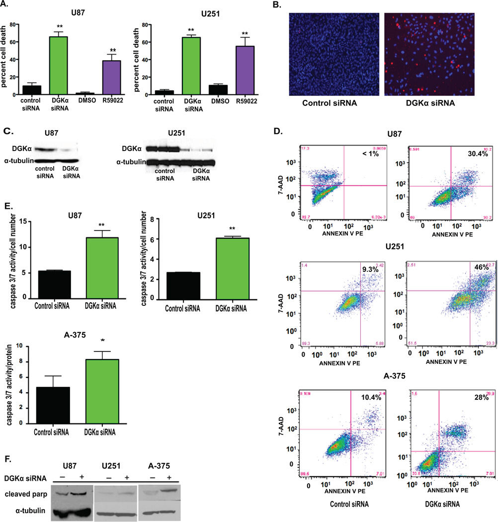
Figure 1. DGKα inhibition causes toxicity in glioblastoma and melanoma cells.
A, DGKα knockdown was assessed in GBM cells U87 and U251 via transfection with either control or DGKα siRNA and inhibition via treatment with DMSO (v:v) or R59022 at 10 µM. Percent cell death was evaluated after 4 days. B, To visualize cell death changes after DGKα knockdown, U251 GBM cells were stained with Hoechst and propidium iodide. C, An immunoblot was used to verify transfection efficiency with siRNA in U87 and U251 cell lysates at 72 hours with α-tubulin as control. D, FACS analysis was performed on both U87, U251, and A-375 cell lines showing an increase in Annexin V-stained cells after DGKα knockdown. E, Caspase 3/7 activity was measured 36–72 hours after DGKα knockdown in U87, U251, and A-375 melanoma cells. F, Protein levels of cleaved PARP were also increased in U251, A-375, and U87 cells after DGKα silencing. (*, P<0.05 and **, P<0.01, Student t test)

