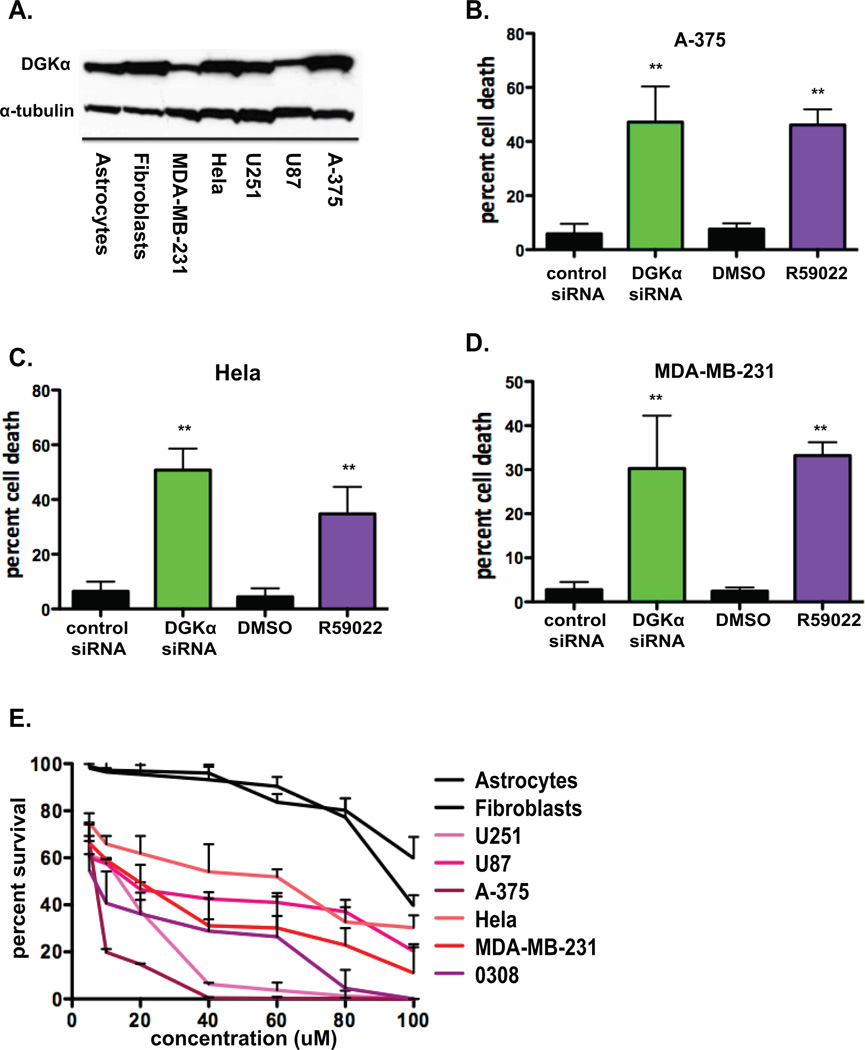
Figure 6. Attenuation of DGKα activity has preferential toxicity for cancer cell line.
A, Basal DGKα levels were evaluated by immunoblot in normal human cell lines and various cancer cell lines, with α-tubulin shown as loading control. Cell toxicity was assessed by cell counts/trypan blue in B, A-375 (melanoma), C, HeLa (cervical cancer), and D, MDA-MB-231 (breast cancer) lines 4 days after DGKα knockdown or treatment with R59022 at 10 µM or DMSO vehicle. E, Dose response curves were generated for astrocytes, fibroblasts, U251, U87, A-375, HeLa, MDA-MB-231, and 0308 (GBM stem cell) cells by cell counts and normalized for DMSO (v:v) at 5, 10, 20, 40, 60, 80, and 100 µM R59022 after 4 days of treatment. (*, P<0.05 and **, P<0.01 Student t test)

