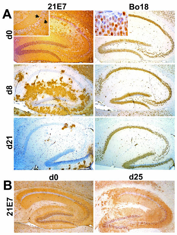
FIG. 4.
Age-dependent BDV resistance of dentate gyrus neurons in Neuro-N-44 mice. (A) Transgenic Neuro-N-44 animals were infected with mouse-adapted BDV strain H215 at the indicated ages (d0, day 0; d8, day 8; d21, day 21). Animals were sacrificed 5 weeks postinfection. Brain hemispheres were paraffin embedded, and 8-μm-thick sections were stained with anti-P MAb 21E7 (left panels) to visualize virus-infected cells or with MAb Bo18 (right panels) to visualize transgenically expressed N. Note the high number of infected neurons in the dentate gyrus of Neuro-N animals infected as newborns. The insert in the upper left panel shows a higher-magnification image of the CA3 region; arrows point to rare infected pyramidal CA3 neurons. The insert in the upper right panel shows a higher-magnification image of BDV-infected dentate gyrus neurons expressing transgenic N. Note the punctate staining pattern in most granular neurons, indicating a redistribution of transgenic N in infected cells. (B) Nontransgenic littermates were infected as newborns (left panel) or at the age of 25 days (right panel) and sacrificed 5 weeks postinfection. Brains were processed as described for panel A, and 8-μm-thick paraffin sections were stained with anti-P MAb 21E7 to visualize infected cells.

