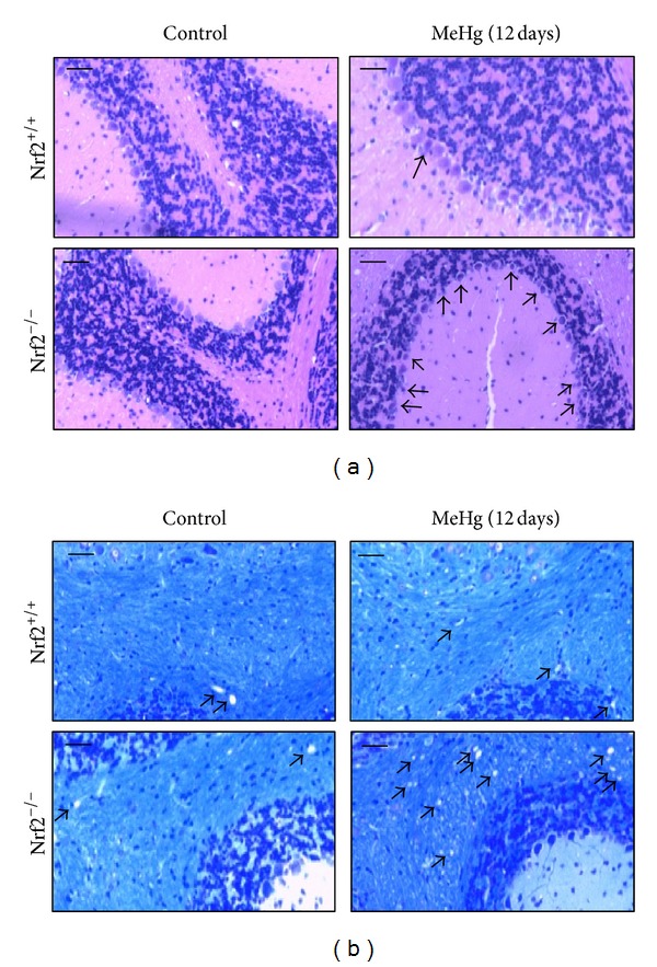
Figure 6.

Pathological observations of brains from Nrf2+/+ or Nrf2−/− mice given MeHg. Nrf2+/+ or Nrf2−/− mice were orally administrated MeHg (5 mg kg−1 day−1) for 12 days, and pathophysiological changes in the cerebellum were observed. Bar = 50 μm. (a) Photographs of the cerebellar granule cell layer (HE stain). Arrows indicate the MeHg-induced degeneration of Purkinje cells. (b) Photographs of the cerebellum medulla (KB stain). Arrows indicate the MeHg-induced vacuolar degeneration.
