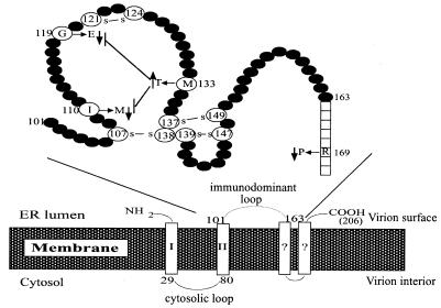
FIG. 1.
Proposed topology of HBV small envelope protein. The lower panel depicts the 206-amino-acid small envelope protein traversing the ER membrane, probably four times. The major cytosolic loop (residues 29 to 80) and luminal loop (residues 101 to 163) are shown. In secreted virions, the luminal loop turns up on the viral surface and is called the immunodominant loop. Its detailed structure, as described by Wallace and Carman (29), is shown above. We have identified in this study four naturally occurring mutations in the S domain that enhance or impair virion secretion. Note that the M133T mutation can suppress the inhibitory effect of G119E and I110M mutations on virion secretion.

