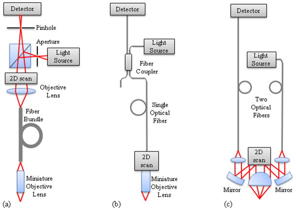
Figure 2.
Fiber confocal microendoscope configurations. (a) Proximally scanned microendoscope using a fiber bundle for illumination and detection. The beam is focused to a point and scanned in two dimensions or to a line and scanned in one dimension at the proximal face of the fiber bundle. (b) Distally scanned microendoscope using a single optical fiber for illumination and detection. Scanning can be accomplished by mechanically scanning the fiber, mechanically scanning the fiber and the miniature lens, scanning the beam with a MEMS mirror, or spectrally encoding one axis and mechanically scanning in the second axis. (c) Distally scanned microendoscope using two optical fibers, one for illumination and one for detection, in a dual-axes confocal configuration with a MEMS scanning mirror.

