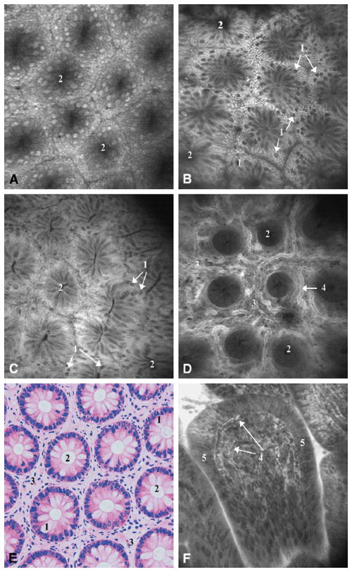
Figure 3.
In vivo endomicroscopic imaging of the normal colon using Optiscan/Pentax (A) first prototype with 6 mm outer diameter and (B–D, F) second prototype with 5 mm outer diameter and integrated into the Pentax EC3870K video-endoscope. Tissue stained with topical application of acriflavin (A–B) and intravenous fluorescein (C–D). (A) Rectal mucosa; (B) descending colon mucosa; (C) cecum; (D) deeper layers of the lamina propria showing microvasculature in the descending colon; (F) terminal ileum. (E) Hematoxylin and eosin stained tissue section cut parallel to the tissue surface for comparison to en face confocal images. 500×500 μm field of view for all images. Reprinted from Gastrointestinal Endoscopy, 62(5), Polglase et al., p. 686–695, Copyright 2005, with permission from Elsevier.99

