
Fig 1. Induction of Th1 cells and Th17 cells from human CD4 T cells.
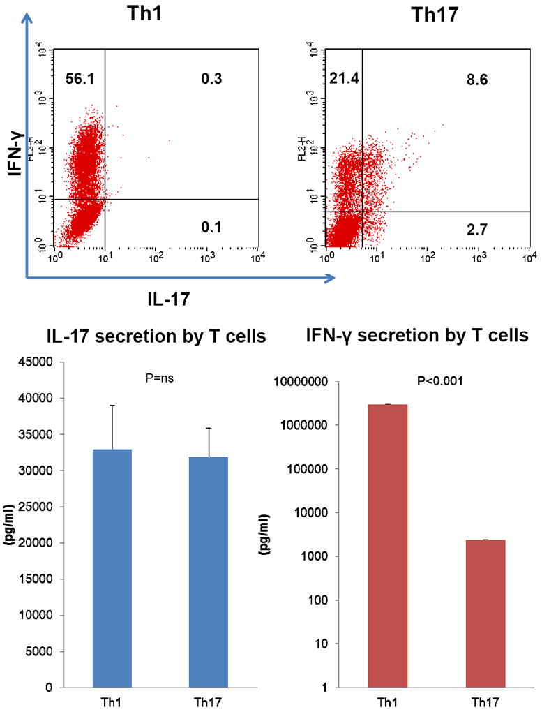
Human CD4 T cells were cultured for 5 days either under Th1 or Th17 polarizing conditions. Following FcR blocking, surface staining of CD4, and intracellular staining of IL-17 and IFN-γ, IL-17 (horizontal axis) versus IFN-γ expression (vertical axis) in the CD4(+) population was assessed by flow cytometry analysis. Representative data of three independent experiments is shown (Fig 1 upper panels). IFN-γ and IL-17 in both Th1 and Th17 polarizing cell culture supernatants were measured by ELISA (Fig 1 lower panels).