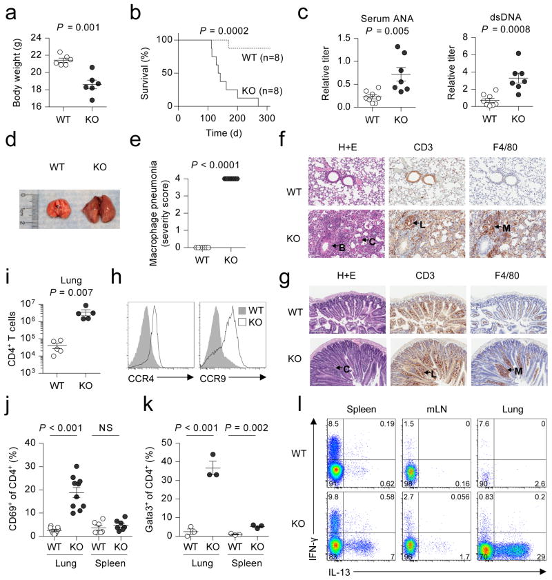
Figure 1. Spontaneous lethal inflammation in Bach2 knockout animals.
a,b, Body weight at three months of age (a) and survival (b) of Bach2 knockout (KO) and wildtype (WT) littermate females. c, Titer of anti-dsDNA antibodies and anti-nuclear antibodies (ANA) in the sera of WT and KO animals. d, Gross morphology of lungs from WT and KO mice. e, Histopathology scoring of lung tissue from WT and KO mice (n = 7 per group). f, Haematoxylin and eosin (H+E) and immunohistochemical (IHC) stains of WT and KO lung tissue with hypertrophy of bronchial epithelium (B), eosinophilic crystals (C), perivascular lymphocytic infiltration (L) and macrophage infiltration (M). g, H+E and IHC stains of small intestinal tissue with hypertrophic crypts (C), lymphocytic infiltration (L) and macrophage infiltration (M). h, Expression of CCR4 and CCR9 on the surface of splenic CD4+ T cells. i, Quantification of CD4+ T cells in lungs of WT and KO animals. j, k, Percentage of CD4+ T cells expressing CD69 (j) and Gata3 (k) in the lungs and spleen. l, Flow cytometry of IFN-γ and IL-13 expression by CD4+ T cells from spleen, mLN and lungs. Mice were analyzed at 3 months of age unless otherwise specified. Data are representative of ≥2 independent experiments with ≥3 mice per genotype. Error bars s.e.m.; P values (Student’s t-test).

