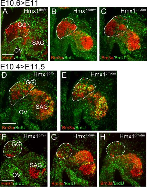
Figure 7. Timing of cell cycle exit in the geniculate ganglion.
Cumulative BrdU labeling was performed in two litters of embryos, beginning early and late on gestational day E10.5 (designated “E10.4” and “E10.6”, see text). (A–C) In the later-labeled embryos, nearly all Hmx1-expressing neurons in the geniculate ganglion (circled) have exited the cell cycle by the start of the labeling period in both control and Hmx1dm/dm embryos. (D,E) In the dorsal geniculate ganglion (circled) of earlier-labeled embryos, some geniculate neurons are still undergoing (a presumably final round) of DNA synthesis at the onset of labeling. A similar fraction of BrdU-positive neurons are observed in the geniculate ganglion of control and Hmx1dm/dm embryos. (F–H) In the ventral geniculate ganglion (circled) of earlier-labeled embryos, only a few Hmx1-expressing somatosensory neurons are present; most of the ganglion at this level consists of Hmx1-negative viscerosensory neurons. Hmx1 neurons at this level have mostly exited the cell cycle by the stage of labeling. Hmx1 expression also identifies a population of early-born neurons in the statoacoustic ganglion. Neither population shows altered incorporation of BrdU in Hmx1dm/dm embryos. GG, geniculate ganglion; OV, otic vesicle; SAG, statoacoustic ganglion. Scale, 100μm.

