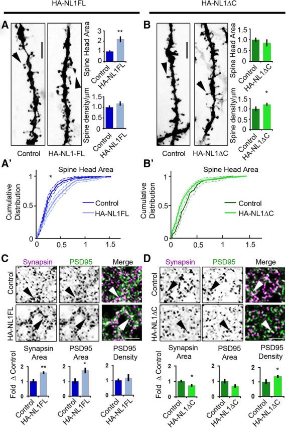
Figure 2.

NL1 intracellular signaling regulates the morphological characteristics of spines and synapses in SLM. A, B, Representative images of dendritic spine segments labeled with DiI of control versus HA-NL1FL mice (A) and control versus HA-NL1ΔC mice (B). Scale bar, 2.5 μm. The mean spine head area was increased for only the HA-NL1FL mice, while spine density was only increased in the HA-NL1ΔC mice (p < 0.05, Student's t test, n = 36 pairs). A′, B′, Cumulative distributions with confidence intervals of spine head sizes across 36 dendritic segments from each transgenic group compared with their controls. Significant differences in the distributions were tested with the Kolmogorov–Smirnov test, p < 0.00001 for controls versus HA-NL1FL mice and p = 0.077 for controls versus HA-NL1ΔC mice. C, D, Representative images and quantification of Synapsin I and PSD-95-positive puncta characteristics of controls versus HA-NL1FL mice (C) and controls versus HA-NL1ΔC mice (D). Areas positive for immunostaining are black. The merge image is shown in color, with Synapsin I in magenta, PSD-95 in green and areas of overlap appearing white. Arrows highlight Synapsin I and PSD-95 colocalization. Scale bar, 2.5 μm. All data are shown mean ± SEM. Student's t test performed in all cases unless otherwise noted, *p < 0.05, **p < 0.01, n = 4 pairs (4 double positive transgenics in each group or 4 mixed single transgenic controls).
