
Figure 4.
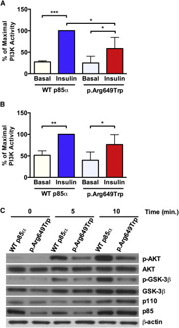
The p85α p.Arg649Trp Substitution Leads to Impaired Insulin Signaling in Primary Human Fibroblasts
(A and B) PI3K activity was evaluated in (A) anti-phosphotyrosine (PY) or (B) anti-IRS-1 immunoprecipitates in WT (normal) and p.Arg649Trp human fibroblast cell lines before and after insulin stimulation (10 mM) for 10 min. Data are expressed as a percentage of maximal stimulation, and p values were calculated by ordinary one-way ANOVA testing (∗p < 0.05, ∗∗∗p < 0.001) (n = 3, data are represented as mean ± SEM).
(C) Immunoblot analysis of lysates obtained from WT (normal) and p.Arg649Trp fibroblasts before and after insulin stimulation was performed with the indicated antibodies. Representative immunoblots are shown.