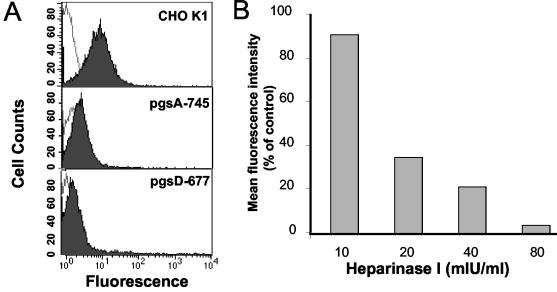
FIG. 4.
Binding of Gs to proteoglycan-deficient CHO cells and effect of heparinase treatment on Gs attachment. (A) Twenty microliters of purified Gs (shown in Fig. 1) was incubated with wild-type CHO K1 cells or with the proteoglycan-deficient cell mutants pgsA-745 or pgsD-677. The attachment of Gs to each cell type was measured by flow cytometry, as indicated in Materials and Methods (shaded histograms). The unshaded histograms correspond to cells incubated without Gs. (B) HEp-2 cells (3 × 10⁵) were incubated for 1 h at 37°C with the indicated amounts of heparinase I before 20 μl of Gs was added to assess attachment by flow cytometry. The results are expressed as the percentage of mean fluorescence intensity relative to that of heparinase mock-treated cells after subtraction of background values (without added Gs).

