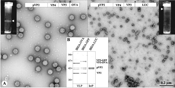
FIG. 2.
Analysis of the structures produced by BacΔIBDA-OVA and BacΔIBDA-LUC. (A) Electron microscopy of the samples purified by CsCl gradient centrifugation. The gradients were illuminated and photographed (inserts). (Left) BacΔIBDA-OVA. (Right) BacΔIBDA-LUC. Material collected from the band present in the gradients was negatively stained with 2% uranyl acetate. Large numbers of VLP-OVA proteins were observed, whereas small irregular particles were obtained with BacΔIBDA-LUC. (B) Polypeptide identification. SDS-PAGE analysis and Coomassie blue staining of the purified VLP-OVA and VLP-GFP and of the irregular particles (IrrP) collected from the IBDA-LUC band. Protein assignment was carried out by tryptic digestion and MALDI-TOF analysis. VLP-OVA proteins were found containing VP3-OVA, VP2, and small amounts of the pVP2 and pVP2 cleaved forms. The relative molecular weights (in thousands), determined by reference to marker proteins, are indicated on the left.

