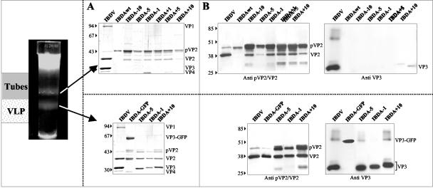
FIG. 5.
Biochemical analysis of the structures expressed by the recombinant baculoviruses expressing IBDA polyproteins modified at the C terminus of VP3. Material extracted from each band shown in Fig. 4 was analyzed by SDS-PAGE followed by Coomassie blue staining (A) or Western blot analyses with an anti-pVP2/VP2 antibody or with an anti-VP3 antibody (B). The upper bands, the tubes, mainly contain pVP2, whereas the lower bands, the VLPs, contain VP2 and VP3. The relative molecular weights (in thousands), determined by reference to marker proteins, are indicated on the left.

