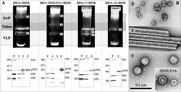
FIG. 7.
Analysis of the structures produced by coexpression of the VP1 polymerase with the IBDA polyprotein and the mutated forms IBDA-DEDLE5A, IBDA − 5, and IBDA − 10. (A, upper panel) After 18 h of centrifugation in a CsCl density of 1.30 at 35,000 rpm, the gradients were illuminated and photographed. The visualized bands were numbered (1 to 3) according to the assemblies they were shown to contain (see panel B). (A, lower panel) SDS-PAGE and Coomassie blue staining of IBDV virions (V) and of the material present in the different bands. Protein assignment was carried out by trypsin digestion and MALDI-TOF analysis. The presence of VP1 was thus ascertained in the different types of VLPs. The relative molecular weights (in thousands), determined by reference to marker proteins, are indicated on the left. IrrP, irregular proteins. (B) Material collected from the band present in the gradients was negatively stained with 2% uranyl acetate. Large numbers of VLPs in band 1, tubes in band 2, and irregular particles in band 3 were observed. For IBDA-DEDLE5A, disk-like structures were also observed. Due to the small amount of material in band 1′, it could not be differentiated from band 1. Note that with the DEDLE5A mutant, small capsids with a different geometry were identified.

