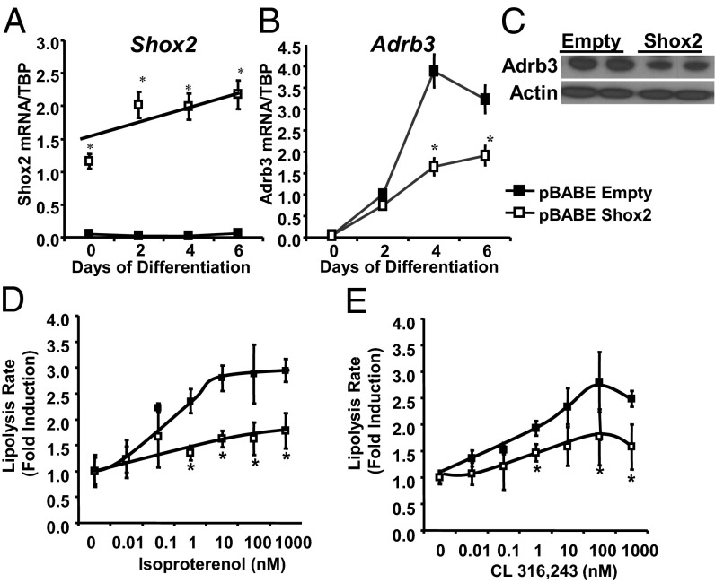
Fig. 5.
Overexpression of Shox2 decreases lipolysis and Adrb3 expression. (A) Expression level of Shox2 mRNA was compared by qPCR between C3H10T1/2 cells stably transfected with pBABE-Shox2 and pBABE-Empty (control) during adipocyte differentiation. Data are mean ± SEM of triplicate samples and the experiment was repeated three times. *P < 0.05 for all panels. (B) Expression level of Adrb3 mRNA was compared by qPCR between C3H10T1/2 cells stably transfected with pBABE-Shox2 and pBABE-Empty (control) during adipocyte differentiation. Data are shown as mean ± SEM of triplicate samples and repeated three times. (C) Western blot of Adrb3 from protein extracts from pBABE-Shox2 and pBABE-Empty C3H10T1/2 adipocytes after 6 d of differentiation. Western blot for actin was used as a loading control. (D) Lipolysis rates after stimulation with isoproterenol as measured by glycerol release from pBABE-Shox2 and pBABE-Empty C3H10T1/2 adipocytes after 8 d of differentiation. Data are graphed as fold induction over basal lipolytic rate ± SEM of three replicates. The entire experiment was repeated twice. (E) Lipolysis rates after stimulation with CL 316,243 as measured by glycerol release from pBABE-Shox2 and pBABE-Empty C3H10T1/2 adipocytes after 8 d of differentiation. Data are shown as graphed as fold induction over basal lipolytic rate ± SEM of three replicates. The entire experiment was repeated twice.

