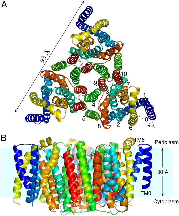
Fig. 1.
Overall view of trimeric YfkE structure. (A) Cartoon representation of an YfkE trimer viewed from the periplasmic side of the membrane with TMs 0–10 colored in rainbow (from blue of TM0 to red of TM10). The pseudo twofold symmetry is highlighted as the dashed line. (B) View from the membrane plane. Hydrophobic membrane area (cyan) is estimated based on multiple tryptophan residue positions near TM termini.

