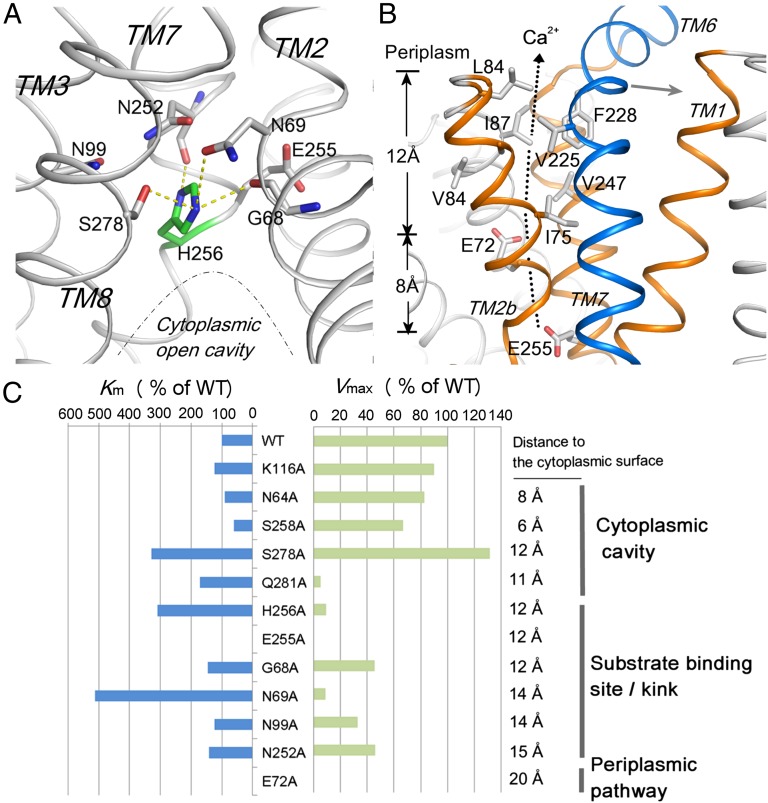
Fig. 4.
Ca2+-binding site and periplasmic closed tunnel conformation. (A) Oxygen umbrella conformation formed by the residues (gray sticks) from the α-repeat helices in the middle of the membrane. The residue H256 (green) is stabilized under the umbrella by hydrogen bond interactions (yellow dash lines). (B) Periplasmic funnel formed by TMs 1, 2b, and 7a (orange) is blocked by TM6 (blue) at the periplasmic exit. The hydrophobic residues along the closed periplasmic tunnel are depicted as sticks. (C) Km (blue bar) and Vmax (green bar) shifts of the YfkE mutants compared with that of wild type. Their position in the Ca2+-translocation pathway and their distance to the cytoplasmic surface are marked.

