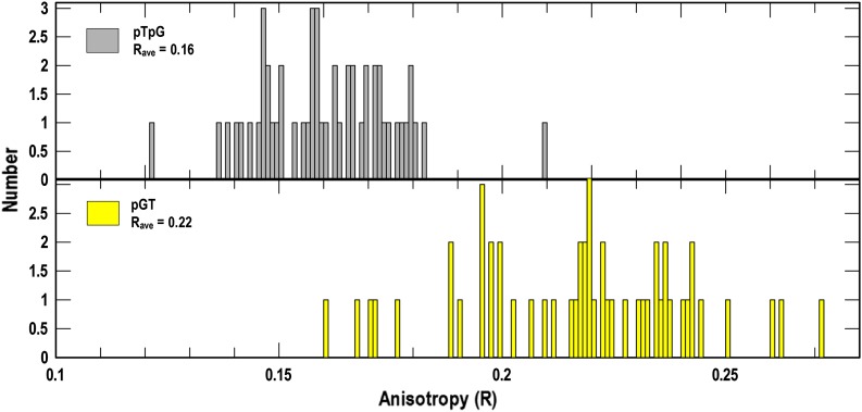
Fig. 2.
Fluorescence anisotropy measurements of bacteria expressing GFP. E. coli BN1071/pTpG (Upper) or /pGT (Lower) (36), which produce cytoplasmic GFP or membrane-associated GFP-TonB, respectively, was grown in MOPS medium and subjected to fluorescence microscopy. We recorded anisotropy (R) of GFP in the two cells for ∼50 measurements from each construct. The resulting histograms revealed a higher R-value, reflecting less rapid motion, for GFP fused to the N-terminus of TonB, resident in the IM bilayer.

