Fig. 1.
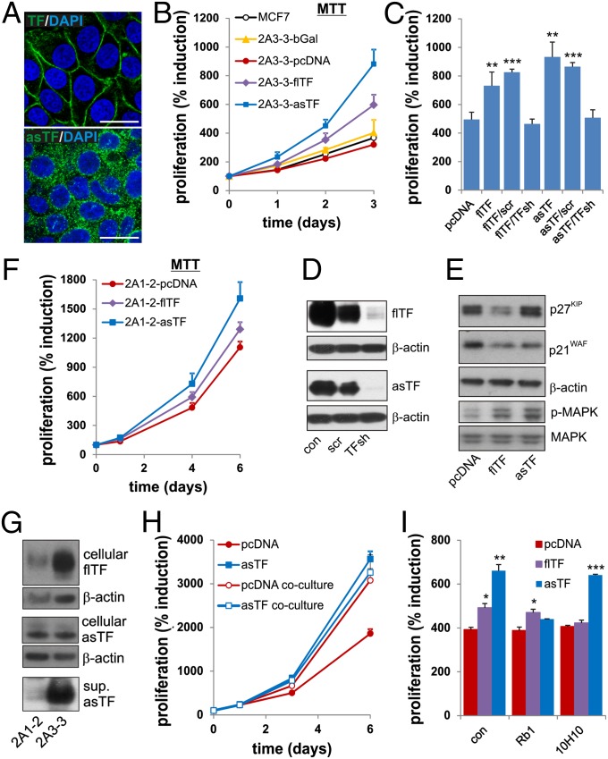
asTF expression induces cancer cell proliferation. (A) FRT cells (clone 2A3-3) received flTF or asTF cDNA by homologous recombination. Localization of flTF (Upper) and asTF (Lower) was assessed by confocal microscopy using specific antibodies [monoclonal flTF-specific Ab 4509 and polyclonal asTF-specific Ab, as described previously (23)]. (Scale bars, 25 μm.) (B) Proliferation of 2A3-3 cells containing the cDNAs indicated was monitored using MTT assay. (C) 2A3-3-flTF or 2A3-3-asTF were transduced with TF-specific or control shRNA. Cells were subjected to MTT assay 3 d after the start of the experiment. (D) flTF and asTF expression in 2A3-3 cells transduced with TF-specific or control shRNA constructs. (E) Expression of cell cycle inhibitors and MAP kinase phosphorylation was assessed in 2A3-3 cell lines. (F) Cell line (2A1-2) harboring an FRT site at a transcriptionally less active region received empty vector, flTF or asTF cDNA and proliferation was monitored using MTT assay. (G) Cell-associated and secreted levels of asTF and flTF in 2A3-3 and 2A1-2 cells. (H) pcDNA cells and asTF-expressing cells were grown separately, or in adjoining 12-well plates containing an open port, allowing asTF diffusion to control cells. Proliferation was monitored using MTT assay. (I) 2A3-3-pcDNA, 2A3-3-flTF, or 2A3-3-asTF cells were treated with 50 μg/mL flTF-signaling blocking antibody (10H10) or asTF-blocking antibody (RabMab1, Rb1), proliferation was assessed 3 d later using MTT assay. *P < 0.05, **P = 0.01, and ***P = 0.001.

