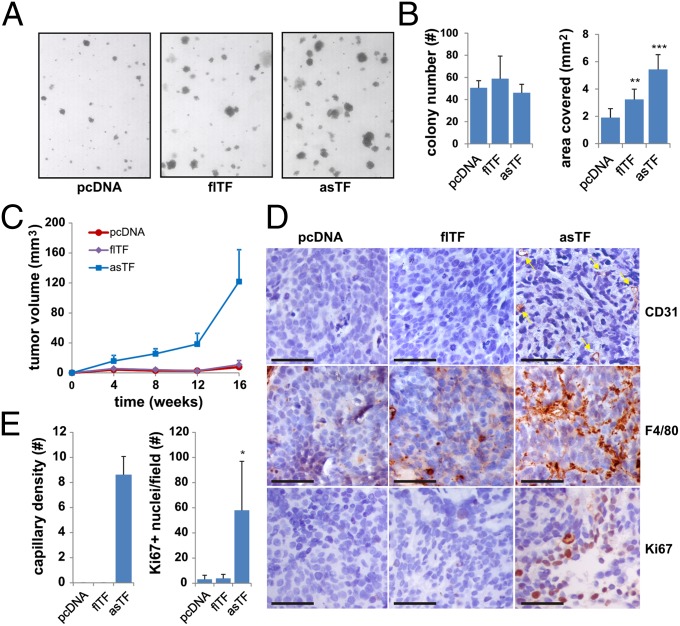
Fig. 4.
TF variant-induced transformation and tumor growth. (A) 2A3-3-pcDNA, flTF or asTF cells were seeded in soft agar and allowed to grow for 14 d. Images were captured and colony number per area covered determined using ImageJ (B). (C) 2A3-3-pcDNA, flTF or asTF cells were injected into the mammary fat pad of NOD-SCID mice, and tumor growth assessed for 16 wk. Mean and SEM are shown. (D) Tumors were analyzed by immunohistochemistry to assess vascular density (CD31), macrophage infiltration (Mac3), and proliferation rate (Ki67). (Scale bars, 50 μm.) (E) Quantification of vascular structures per view field. *P < 0.05, **P = 0.01, and ***P = 0.001.

