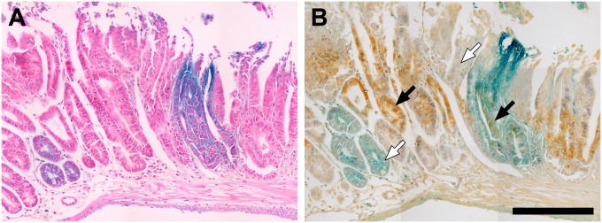
Fig. 4.

Polyclonal tumors are composed of blue and white neoplastic cells. (A and B) To confirm the impression of two pathologists based on hematoxylin and eosin stained sections (A), immunohistochemistry was performed to assess the localization of β-catenin (B). Blue and white cells within the the tumor clearly have β-catenin localized to the nucleus (black arrows), which is a marker for neoplastic transformation, whereas histologically normal cells of either color do not (white arrows). (Note that 4A and 4B are composites to create the full images.)
