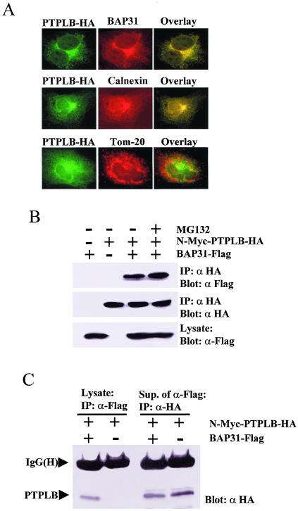
FIG. 5.
PTPLB associates and colocalizes with BAP31 in the ER. (A) Cos-1 cells grown on coverslips were transfected with PTPLB-HA for 18 h, followed by the addition of MG132 to the medium for 4 h, and the cells were double stained with anti-HA antibody (green) and either anti-BAP31 (top panel), anti-calnexin (ER marker, middle panel), or anti-TOM20 (mitochondrion marker, bottom panel) (red), and images were visualized in the red, green, and yellow (overlay) channels. (B) Association of PTPLB with BAP31 in Cos-1 cells. Cos-1 cells were transfected with cDNAs coding for N-Myc-PTPLB-HA and/or BAP31-Flag as indicated. Anti-HA immunoprecipitates (IP) were recovered and resolved by SDS-PAGE, and immunoblots were probed with anti-Flag (top panel) or anti-HA (middle panel). Equivalent samples from cell lysates were immunoblotted with anti-Flag (bottom panel). (C) Small amounts of PTPLB associate with BAP31 in Cos-1 cells. Cos-1 cells were transfected with cDNAs coding for N-Myc-PTPLB-HA with or without BAP31-Flag as indicated. Anti-Flag immunoprecipitates (IP) were recovered and resolved by SDS-PAGE, and immunoblots were probed with anti-HA (lanes 1 and 2). Subsequently, the remaining PTPLB molecules were collected from the BAP31-depleted supernatants with an anti-HA antibody, and the immunoblots were probed with anti-HA (lanes 3 and 4).

